Medical and diagnostic work by the staff of the Department of Maxillofacial Surgery of the Belarusian State Medical University (have the highest (22) and first (4) qualifying medical category) is carried out on the basis of inpatient departments of maxillofacial surgery 11 of the City Clinical Hospital of Minsk, Republican Scientific and Practical Center of Oncology and Medical Radiology named after I. N.N. Aleksandrov, surgical dental offices of the RKSP, 3, 4, 11, 13 dental and 2, 4, 8, 10, 17, 25, 30, 31 general polyclinics in Minsk, the Minsk Clinical Diagnostic Center, the teaching and learning center.
At these bases in 2016, the staff of the department carried out more than 500 inpatient (including 50 high-tech and 31 complex surgical interventions), more than 800 outpatient emergency and planned surgical interventions and more than 6,000 tooth extractions, consulted more than 10,000 patients with various congenital and acquired pathologies face and neck.
The Department of Oral and Maxillofacial Surgery continues its research work together with the Belarusian State University of Informatics and Radioelectronics. Together with the UE 'STC' LEMT 'BelOMO', a method of using a laser for surgical interventions has been developed and introduced into practical health care.
In 2016, 3 new methods of diagnostics and treatment of pathology of the maxillofacial region were introduced at the Minsk Healthcare Institution (UZ 11 GKB). The main directions of the department's work are the development of innovative methods of surgical interventions on the bones of the midface zone, large salivary glands, neck cysts and bone grafting of the jaws.
In 2016, the staff of the department made 47 emergency visits to medical institutions in Minsk for consultations and providing the necessary specialized care to patients. Participated in the work of the commissions created by orders of the Ministry of Health of the Republic of Belarus and the Health Committee of the Minsk City Executive Committee.
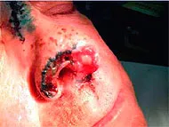
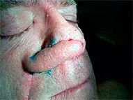
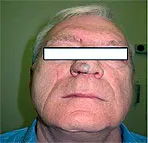
Examples of reconstructive operations
Stages of plastic elimination of a post-traumatic nasal defect with a nasolabial fold flap:
Reconstruction of the alveolar processes of the jaws for dental implantation:
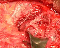
Stages of microsurgical surgery to remove a tumor of the parotid salivary gland using an operating microscope:

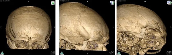
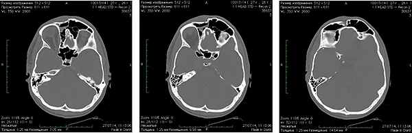
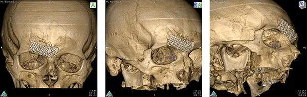
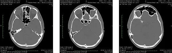
Open reposition of the anterior wall of the frontal sinus, osteosynthesis of the anterior wall of the frontal sinus with a titanium shield
Spiral computed tomography before surgery:
Spiral computed tomography after surgery:
The staff of the department took part in the development and implementation of more than 40 diagnostic, treatment methods, and the use of new instruments and equipment in practical health care.
Basic techniques:
"The use of magnetic resonance computed tomography in the diagnosis of tumors of the face and neck";
"The use of magnetic resonance computed tomography in the diagnosis of pathologies of the large salivary glands";
"Microsurgical technique of organ-preserving operations in pathology of the large salivary glands";
"Sialodochostomy technique for salivary stone disease of the large salivary glands";
"The use of bioactive ceramics based on calcium phosphate for filling the postoperative bone cavities of the jaw bones";
"Application of polymer-composite implantation material" IKVOBAN - Mt1 "for contour plastics in the maxillofacial area";
"Method for the formation of organoplastic material in the elimination of through defects in the oral area";
The use of various designs of titanium maxillofacial implants (standard and individual) for reconstructive operations on the bones of the facial skull;
Optimization of diagnostic methods and predicting the course of purulent-inflammatory processes in the maxillofacial region;
Development of new diagnostic methods and treatment of patients with various surgical pathologies of the large salivary glands (benign tumors, SKB, pyoinflammatory processes), including the improvement and development of organ-preserving operations;
Minimally invasive techniques for removing the midline formations of the neck;
Use of an autobone graft with a cortical layer to eliminate postsequestral osteomyelitic bone defects of the upper jaw;
A method of treating a hypervascular tumor of the skull base;
Complex method for diagnosing salivary stone disease of the submandibular salivary glands;
Restoration of the continuity of the mandibular bone using non-vascularized autograft from the iliac crest;
Application of the SMAS-flap base to the auricle during operations on the parotid gland;
Method of transfocal osteosynthesis using a mesh plate of complex shape;
The technique of sparing incisional biopsy on the oral mucosa in order to verify pathological processes.
Planning of reconstructive operations on the bones of the facial skeleton using stereolithography and performing surgical interventions using reconstructive plates and titanium mesh implants.
Improvement of diagnostic and treatment methods for diseases of the large salivary glands and
neck cysts.